Dr. Ratnav Ratan suggests suitable surgical measures to treat crouch gait in children with
Cerebral Palsy and regain their mobility.
Crouch gait is a common gait abnormality detected in children affected by cerebral palsy. The
crouch gait is a challenging condition with a high rate of recurrence. It is observed that there is
excessive knee flexion in the child.
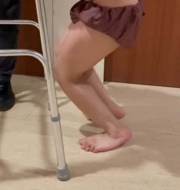
Mobility of a child with Crouch Gait
Crouch gait leads to impairment of the mobility of the child. The gait requires more effort, and
hence, the child tires easily and complains of joint pain. The child puts additional strain and
pressure on the bones, joints, and muscles to move and maintain balance. There is an ongoing
contraction of knees, hips, and ankles, which puts pressure on select muscles continuously, while
some muscles lie unused to a great extent. The child utilizes more energy to move around, which
does limit the extent of activities feasible for the child.
This imbalance in the lower extremities leads to joint pressure and chronic pain. In addition, the
child complains of knee pain with the onset of arthritis as there is an excess burden on the
anterior knee and failure of the extensor mechanism to perform.
In the case of improper management of legs for spasticity, the child may develop more
complications such as
- Inability to walk
- Dislocation of joints
- Weakened hip and knee extensor
- Weak ankle plantar flexors
- Uneven and distorted growth
Identifying the crouch gait
To begin with, the child develops an irregular walking pattern characterized by upward bending
of the ankles, bent knees, and hips. The child assumes a crouching position when walking, and
hence the gait is referred to as a crouch gait.
Conditions that cause crouch gait
Involuntary muscle contractions are known as spasticity, which indicates faulty communication
between the muscles and the brain. As a result, these muscles remain contracted for a long time
as the brain is not in a position to signal them to relax.
A child develops cerebral palsy when their developing brain is damaged; the extent of damage
depends upon the location of the brain damage. Though the brain damage does not extend
further, spasticity can worsen, leading to increased complications in the functioning of the
muscles
In such cases, pediatric orthopedic surgeons suggest a comprehensive surgery plan to resolve this
a progressive condition, while the approach depends upon the patient’s physiologic abnormalities
and anatomic conditions.
Noted pediatric orthopedic surgeon in Delhi and Gurgoan, Dr. Ratnav Ratan, suggests
performing the single-event-multilevel-surgery – SEMLS, the most reliable and preferred
surgery, which is performed on patients suffering from cerebral palsy and spina bifida.
The SEMLS procedure is performed on hips, knees, ankles, and feet to make the required
corrections in the soft tissues and bones. SEMLS implies a single corrective procedure followed
by a consolidated rehabilitation protocol.
The benefits of a SEMLS procedure
- Enhancement of mobility skills
- Preservation of walking abilities or improvising them
- Preservation of joint health and mobility
- Reduce the consumption of oxygen, which means the child will feel more active.
- Better management of muscles and bones that are misaligned
- Better pain management and lessen the intensity of pain in the future.
A child was diagnosed with neurologic conditions that have affected the brain or the spinal cord,
leading to walking abnormalities and gait issues. If left untreated, the child will suffer from
arthritic joints that will be painful and limit the child’s mobility
Following are the components of SEMLS:-
Distal femoral extension osteotomy and patellar tendon shortening
The treating paediatric surgeon performs two surgical procedures, viz., the distal femoral
extension osteotomy and patellar tendon shortening, simultaneously. In the distal osteotomy
procedure, the surgeon removes the anterior wedge from the distal femur to straighten the lower
limb and compensate for the static contracture of the posterior capsule and hamstring muscles.
The knee flexion deformity is also corrected through distal osteotomy. Through the patellar
tendon shortening, the high riding patella is brought down. A plate is used to keep the correct
position of the femur. The healing period is almost six weeks or more.
The child’s gait kinematics improve greatly, especially at the knee joint. Moreover, the improved
condition is maintained for quite a long period of time.
Tendo-Achilles shortening in Cerebral Palsy Children
Tendo-Achilles shortening is performed to reduce the calcaneus deformity of the ankle on weight
bearing.
Hip- Iliopsoas lengthening in Crouch gait
It helps reduce the anterior pelvic tilt.
Semitendinosus transfer to Adductor Magnus
Limited release of Hamstring muscles
Dr. Ratnav Ratan will suggest the use of orthotic devices postoperatively. Braces, splints, and
other orthotic devices are useful in supporting and promoting the desired musculoskeletal
alignment, enabling subtle stretching of spastic muscles, and preventing contractions. With long
term use, once the body and the mind adjust to the desired gait, the child can slowly stop wearing
them and walk properly.
Children with crouch gait and other abnormal walking patterns need consistent management.
Also, their condition is likely to deteriorate further without regular management. Hence, early
detection and treatment is advisable to improve their balance and posture while focusing on
reducing the intensity of pain and pressure on the limbs and joints